Scrotal Ultrasound
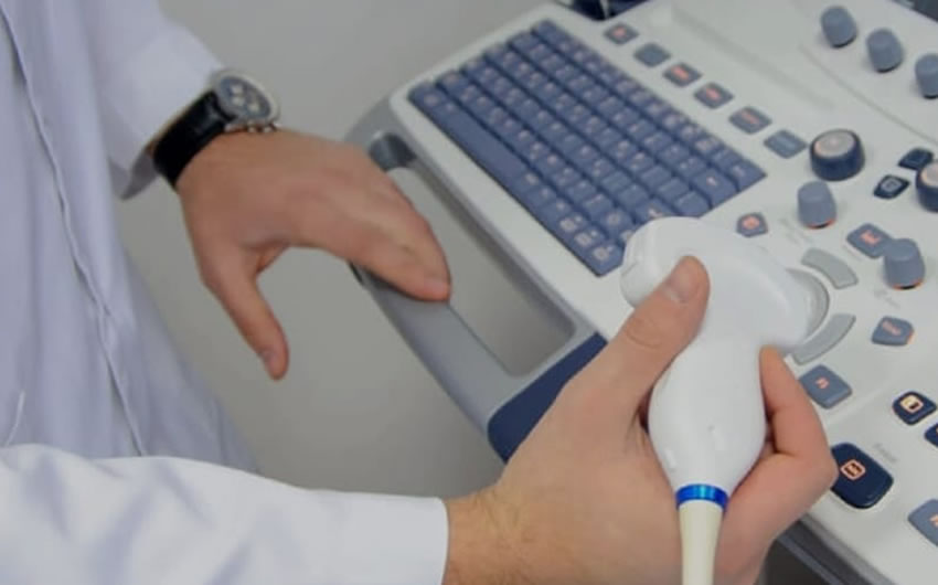
Ultrasound examination is the primary imaging choice for various scrotal conditions. Using a high-frequency linear transducer, we offer superior detail compared to CT or MRI, making it an ideal diagnostic tool for new lumps in the scrotum. Patient comfort, privacy, and dignity are paramount during this intimate examination.
We meticulously examine the testes, epididymides, and scrotal wall, assessing symmetry, echotexture, and echogenicity. We also consider age-related changes and post-vasectomy alterations. Our examination extends to evaluate conditions like varicocele, scrotal masses, suspected testicular torsion, and testicular microlithiasis. Our scrotal ultrasound aims to provide accurate diagnostic insights, and we prioritise patient well-being and comfort throughout the process. In cases where urgent surgical exploration is necessary, our practitioners are trained to identify ultrasound features related to torsion.

For specific management guidelines and follow-up, we work closely with urological surgical colleagues and radiologists, ensuring comprehensive patient care.
